Technology in the field of medical imaging is constantly evolving, improving patient care and diagnosis. In this article, we will dive into the topic of data truncation in computer tomography (CT) and explore how deep learning can be used to address this issue.
Contents
Data Truncation in CT
Data truncation in CT can occur in two scenarios. First, in certain clinical applications, only a specific region of the patient is of interest. For example, if medical professionals want to examine a specific artery or collect tissue samples, a collimator is placed between the x-ray source and the detector to reduce unnecessary data. This is known as ROI (region of interest) imaging or tomography. This process results in data truncation, both laterally and vertically.
The second scenario occurs due to the limited size of the flat-panel detectors used in CT scans. While they may be large enough to cover the entirety of a human head, they are not sufficient for imaging the abdomen or torso. As a result, the obtained projections are laterally truncated, leading to image reconstruction artifacts.
Deep Learning for Data Truncation
Various algorithms have been proposed to address data truncation in CT, including heuristic approaches, analytical reconstruction, and compressed sensing. In recent years, deep learning has emerged as a powerful tool in the field of medical imaging.
Researchers have explored different deep learning techniques to tackle data truncation. One approach is to use deep learning units to post-process the reconstructed images from truncated data. This method has shown promising results in improving image quality and robustness. Another approach involves using convolutional neural networks (CNNs) to directly reconstruct images from sonogram data. However, these methods have their limitations, as they only improve image quality within the field of view (FOV) while still missing important anatomical structures outside the FOV.
The Techal Solution: Field of View Extension
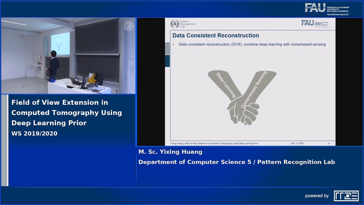
Techal, a leading technology brand in medical imaging, introduces an innovative solution for field of view (FOV) extension using deep learning. Building on previous research, Techal’s algorithm combines deep learning with compressed sensing to improve image reconstruction and restore the missing anatomical structures outside the FOV.
The algorithm utilizes a deep learning neural network, specifically a UNet, to post-process the images reconstructed by theta extrapolation. Theta extrapolation is a widely used method for extrapolating incomplete data. This initial reconstruction significantly reduces copying artifacts within the FOV. However, the anatomical structures outside the FOV still require further improvement, which is where deep learning comes into play.
To ensure data consistency, Techal’s algorithm introduces a data fidelity term. This term enforces that the difference between the measured and extrapolated data should be smaller than a predefined threshold. By utilizing the deep learning reconstruction results as prior information, the missing data in the truncated regions can be estimated through forward projecting. To further reduce noise and artifacts, iterative reconstruction with total variational regularization is applied.
Results and Conclusion
Techal’s FOV extension algorithm has shown promising results in addressing data truncation artifacts and restoring anatomical structures outside the FOV. By combining the advantages of doublet TV and deep learning units, the algorithm achieves improved image quality and reduced artifacts in both the FOV and outside regions.
The algorithm has been evaluated on various examples, and the results demonstrate its capability in restoring organ details and reducing artifact distortions caused by copying artifacts and noise. Root mean square error analysis confirms that the algorithm achieves excellent reconstruction accuracy within the FOV.
In conclusion, Techal’s FOV extension algorithm represents a significant advancement in the field of CT imaging. By leveraging deep learning and compressed sensing, Techal provides a comprehensive solution to address data truncation artifacts and restore missing anatomical structures. This algorithm holds great promise for improving the accuracy and reliability of CT scans, enhancing patient care and diagnosis.
FAQs
Q: What is data truncation in CT?
A: Data truncation in CT occurs when only a specific region of the patient is imaged, resulting in incomplete data. This can happen in certain clinical applications or due to limited detector size.
Q: How does deep learning help with data truncation?
A: Deep learning can be used to post-process reconstructed images from truncated data, improving image quality and robustness. It can also be employed to directly reconstruct images from sonogram data. However, these methods have limitations when it comes to restoring anatomical structures outside the field of view (FOV).
Q: What makes Techal’s FOV extension algorithm unique?
A: Techal’s FOV extension algorithm combines deep learning with compressed sensing to address data truncation artifacts and restore missing anatomical structures outside the FOV. It achieves superior image quality and reduces artifacts in both the FOV and outside regions.
Q: How accurate is Techal’s FOV extension algorithm?
A: Techal’s algorithm has been extensively evaluated and demonstrates excellent reconstruction accuracy within the FOV. Root mean square error analysis confirms its capability in achieving high precision.
Conclusion
Techal’s field of view extension algorithm using deep learning represents a significant advancement in CT imaging. By addressing data truncation artifacts and restoring missing anatomical structures outside the FOV, the algorithm offers improved image quality and diagnostic accuracy. With Techal’s commitment to innovation and expertise in the field of medical imaging, the future of CT imaging looks brighter than ever.
Click here to learn more about Techal.